Category Archives: Uncategorized

Case Vignette – January 2020

CASE VIGNETTE – JANUARY 2020

Non Sequitur is a Real Phenomenon

It can often be accepted that an accident occurred. In addition, an injured party may exhibit significant impairment and loss. What is not always so clear is the presence or otherwise of a link between those two events.

The Latin term “non sequitur” refers to the truth incumbent in both statements, but the complete absence of causation.

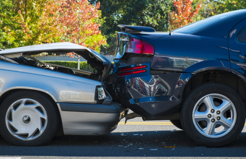
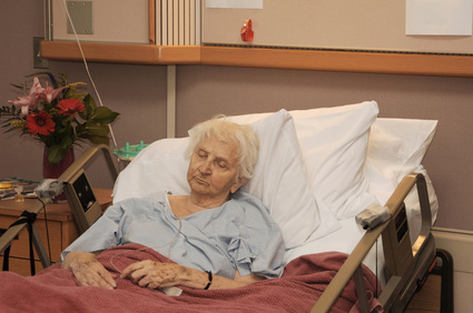
I recently saw a 60 year old male who was a rear seated passenger in a motor vehicle which turned right across the path of oncoming traffic. His driver was obviously at fault but the injuries he sustained remained real.

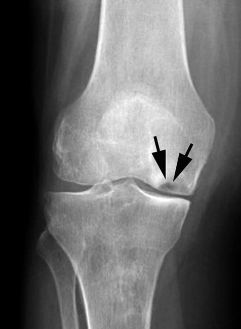
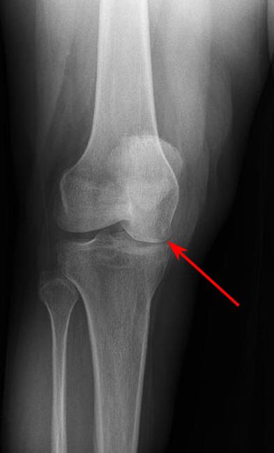
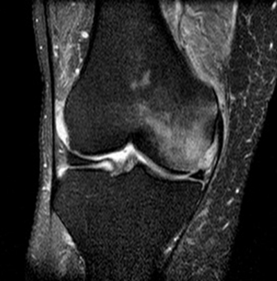
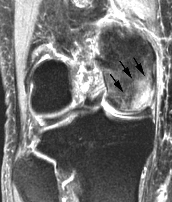
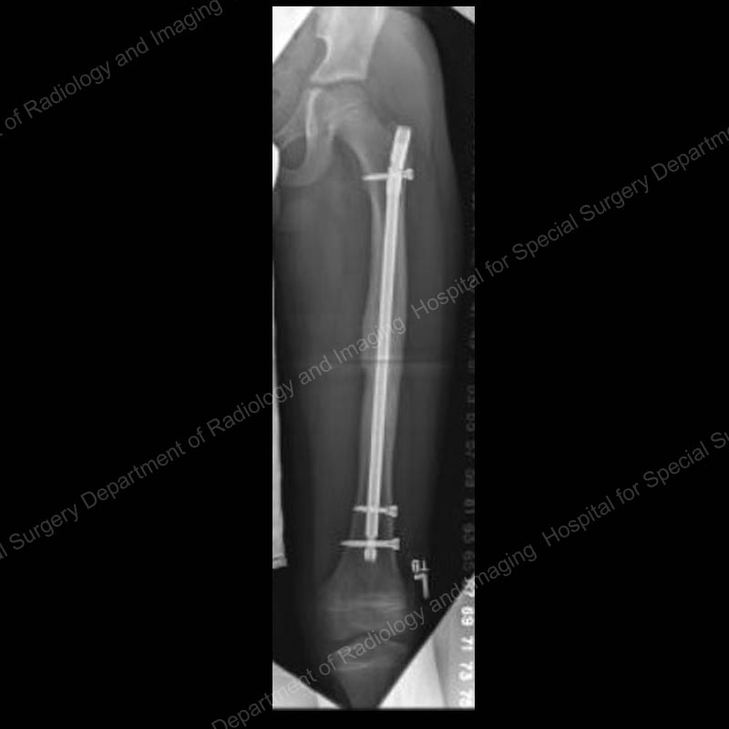
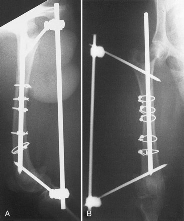
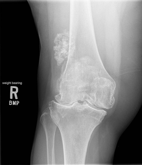
One of the injuries related to his right knee. There was a direct blow over the front of the patella (the knee cap) on the dashboard. This same knee had been subjected to an anterior cruciate ligament reconstruction 20 years previously. If the plaintiff was to be believed, the reconstruction had been an outstanding success and the knee joint had remained asymptomatic up until the time of this road traffic accident.

In assessing his circumstance, it appeared clear that whatever functional loss may have predated the accident as a result of the cruciate ligament reconstruction many years previously, it was not linked with the accident. Conversely, there was some additional loss that could be linked with the accident. The blow to the patella had damaged the joint between the knee cap and the high bone and had given rise to a measurable loss using the AMA 5 Guides.


Whilst the defendant’s team was keen to discount any knee problem as a result of the accident because of this positive past history, the plaintiff and his advisor were not easily dissuaded.

Common sense prevailed, compensation was forthcoming and to my mind, it was fair.

The lesson is important, and sometimes overlooked. Damaged goods can be made worse. A pre-existent functional impairment can be added to. Significant additional impediments can accrue.

It is only with a full history, a thorough clinical examination and a careful analysis that a true apportionment of blame can be identified.

Case Vignette – January 2021

Case Vignette – January 2021

The Metal-on-Metal Debacle

Hip replacements usually involve a metal or ceramic ball articulating with a plastic, ceramic or metal cup, or acetabular component. Articulations between metal/ceramic and plastic or polyethylene usually perform very well and all the more so given recent advances in the manufacture of highly cross-linked polyethylene acetabular components.

Unfortunately, metal femoral heads articulating with metal acetabular components have had a checkered career. The concept is not new although since the year 2000, hundreds of thousands of these large head metal on metal devices were implanted unwittingly in patients by surgeons who knew no better.

Not all of these metal-on-metal articulations were destined for failure but unfortunately, an unacceptably high percentage did.

Revision surgery was almost always required and complex procedures were mandated to deal with the adverse effects of the metal-on-metal debris that was accumulated.

There have been class actions in North America and Europe aimed at achieving recompense for the multitude of patients so affected. In Australia, there have been no successful class actions to date although some may still be brewing.

The difficulty arises in sheeting home the blame. The manufacturers appear to have complied with all of the dictates of the Food and Drug Administration or the Therapeutic Goods Administration in Australia. The surgeons also undertook appropriate preliminary evidence gathering and the warning signs were not present until 2008 or thereabouts. The patients could hardly be blamed at all.

Whilst these large head metal-on-metal devices are now rarely used, it is possible that the legal cesspit continues to bubble along.


Case Vignette – July 2018

CASE VIGNETTE – JULY 2018

Are MRI Scan Examinations Important?

Sometimes!

There is no substitute for the taking of a full history and the performance of a thorough physical examination. A good Orthopaedic Surgeon can usually make a diagnosis in 85% or more cases just with those two useful tools. Plane radiographs will increase the diagnostic accuracy up to the region of 92%. The remaining 8% of difficult diagnoses can usually be secured with additional investigations such as CT scans, MRI scans, bone scans or PET scans.

knee x-ray 5

READ MORE

MRI scan examinations can be very useful. They are best used to analyse soft tissue injuries although can also be used to assess bony or skeletal injuries.

I well recall a patient who was in his fifties. He had been subjected to a knee injury. He was adamant that this particular knee had been completely asymptomatic prior to the subject injury.

When plane radiographs were performed within a few days of the injury, it was found that he had a significant abnormality involving that knee. He suffered with so-called avascular necrosis of the medial femoral condyle. This is an area of death within the bone which forms part of the lower end of the thigh bone and would definitely have been present prior to the injury. It is possible however, although relatively unlikely, that he may have been completely unaware of the condition at a clinical level before the injury. A very careful and thorough scrutiny of his clinical records failed to reveal any evidence that he had ever complained of any problems referable to that knee joint. The benefit of the doubt therefore lay with him.

knee x-ray 6


The defendants argued that the subject injury had simply unmasked this condition and had not really caused any particular damage or harm.

Fortuitously, he had been subjected to an MRI scan examination within three weeks of the subject accident. Not only did the MRI scan examination show these old changes of avascular necrosis involving the medial femoral condyle, but the scan also showed oedema in the bone and the soft tissues in that region. So extensive was the oedema in the soft tissues that it was reasonable to assume that quite considerable forces had been applied to the joint at the time of the subject injury and that at least some of his presentation was due to the injury specifically.

mri 7

This became of considerable importance. Although he had been asymptomatic prior to the injury and it would be reasonable to assume that he may have continued in that asymptomatic state for some considerable time into the future, his clinical course was altered by the injury. Within six months, he had undergone a partial knee replacement. Unfortunately, that intervention failed and within thirteen months, he had undergone a total knee replacement. That intervention was also not particularly successful and given the fact that he was still in his fifties, it was more likely than not that he would eventually require a revision of that total knee replacement.
He had therefore gone from a state of being completely asymptomatic to a circumstance whereby he had been subjected to two major operations yet remained seriously encumbered.

He was successful in his civil suit and the quantum he received was thought by the defendant to be extremely generous. Whilst that may have been the case, had it not been for the MRI scan examination findings, I suspect that he would have received little, if anything.

mri 8

In this situation, the MRI scan examination appeared to be pivotal.

Case Vignette – July 2019

CASE VIGNETTE – JULY 2019

Not Everything Is As It Seems

Orthopaedic surgeons rely heavily upon radiographs to diagnose fractures. Fractures in themselves can be sub-classified in a myriad of ways. In essence however, a fracture refers to a discontinuity in the normal structure of a bone. Bones are living organs and have cortical and cancellous components.

Plane radiographs (and I use the word advisedly) are simply two-dimensional representations of a three-dimensional happening. Considerable overlay will be present on the film and an undisplaced, relatively minor fracture may not initially be visible.

Normal scaphoid

The scaphoid bone (one of the small bones in the wrist) is a classic example. The scaphoid is about the size and shape of a cashew nut. It is vulnerable to fractures through the waist or the mid-section, especially in patients who sustain a fall on an outstretched hand or wrist.

The patient will almost invariably complain of pain in the so-called anatomical snuffbox (the small space just at the base of the thumb), but the initial x-ray or radiograph may show nothing untoward.

a00012f04

Part of the biology of fracture healing is initiated by so-called hyperaemia or an increase in blood flow with resorption of radio-opaque minerals at the fracture edges. Once this process has been in train for ten to fourteen days or so, some of the minerals are leached from the fracture zone and the region appears darker on a subsequent x-ray or radiograph. The diagnosis is then easier to make. As the healing process continues, new mineral is deposited, the area becomes more radio-opaque or white and the diagnosis is obvious.

The real trick here is to have a high index of clinical suspicion. If the patient does have a history of having fallen on an outstretched hand or wrist, does have considerable pain localised to the anatomical snuffbox and yet has a normal radiograph, it is prudent to immobilise the wrist as though a scaphoid fracture was present. The cast can then be removed two weeks later, a new radiograph performed and the underlying presence of a scaphoid fracture can either be confirmed or excluded.

scaphoidnonunion1

Failure to have this suspicion at the outset, and the failure to immobilise the wrist immediately, increase the risk of delayed or non-union with obvious ramifications.


Case Vignette – July 2020

CASE VIGNETTE – JULY 2020

How Valuable are Photographs of the Accident Site in Assessing Injuries Sustained?

They are relatively useful. I use that adjective “relatively” advisedly. I understand that extremes can occur. For example, some passengers may escape unscathed despite a vehicle being completely wrecked. Conversely, what might appear to be very minor damage to a vehicle could result in some significant physical injury. In general terms though, the injury spectrum and severity increase proportionately to vehicle damage.

I recall a case some years ago. A female driver’s motor vehicle had been struck from the rear by another vehicle. The total extent of the damage extended to one plastic indicator light cover. Nothing more. The indicator light cover was replaced for $84.50. You would expect that the forces applied to the female driver’s car were relatively minor.

Clinical and radiological investigations confirmed that she had evidence of a fracture in her thoracic spine and that her 14-week-old foetus was dead in utero. Through a lack of careful analysis, both of these findings were attributed directly and causally to the subject road traffic accident. The offending driver, believing that he was responsible for the death of the foetus, subsequently took his own life.

It transpired that the thoracic fracture was old. It had resulted from a fall from a horse over a decade previously. In addition, although the foetus had died in utero, it was evident subsequently that the intrauterine death had occurred some six weeks prior to the road traffic accident. In other words, it was a so-called missed abortion.

This was a true tragedy. There was no causal link between this minor road traffic accident and the sequence of events that followed. Instead, the imprudent carriage of the case resulted in a totally unnecessary loss of life.

Whilst not necessarily accurate or predictive of injury severity, the extent of a vehicle damage does play some part in the final analysis.

Case Vignette – July 2021

Case Vignette – July 2021

Osteoporosis of Pregnancy

Osteoporosis refers to demineralisation of the collagen structure in bone. It presents clinically as fractures caused by minimal force and radiographically by bones that are far less radio-dense than normal. Whilst the condition is often associated with age, and in particular in females, it can also rarely be associated with the third trimester of pregnancy in quite young women. Mothers at or about the age of 30 years appear to be particularly vulnerable and it is during the third trimester that hip pain (either unilateral or bilateral) is noted.

The diagnosis initially requires a high index of suspicion and this is not always present with all obstetricians. The diagnosis is best made with an MRI scan examination and the treatment modalities will differ according to the extent of the lesion that is identified.

Ideally, the condition is identified in its earliest phases, before a fracture of the femoral neck occurs and before anything more than rest and observation are required.

Sometimes, if the diagnosis is delayed, a fracture of the femoral neck may ensue. If that fracture is incomplete or undisplaced, it can be managed relatively easily with internal fixation. This is an orthopaedic endeavour that is not usually very difficult. Most fractures will then go on to sound osseous union with little or no long-term sequelae.

The most undesirable state however is when the diagnosis is delayed, a fracture occurs, the fracture becomes displaced and the risk of non-union, delayed union or avascular necrosis of the femoral head increases exponentially. If the fracture is displaced, many of these patients will eventually require a total hip replacement.

Whilst the hip replacement will solve the problem initially, it is an undesirable operative intervention in somebody who is in their early thirties. Hip replacements have a finite life span and theoretically, 10% of those patients will require a revision by the time they are in their early fifties. Of those, a further 20% will require a second revision in their seventies. Even a third revision may be required in the patient’s eighties, given actuarial longitudinal life studies that exist today.

It is important therefore that all clinicians involved in obstetric care have at least a high index of suspicion for this unusual and uncommon condition. Early diagnosis, proper orthopaedic care and judicious rehabilitation can effect an excellent outcome. If any or all of those elements are absent, the outcome can be far from desirable.

Case Vignette – June 2019

CASE VIGNETTE – JUNE 2019

More Than One Way To Skin A Cat

I recently saw a patient who had a long spiral fracture of his femur or thigh bone. It had been managed operatively with the insertion of a rod through the centre of the bone with interlocking screws above and below the fracture.

The rod acts to provide longitudinal stability for the fracture and prevents angulation at the fracture site. The interlocking screws above and below provide rotary stability and also prevent the fracture overriding or “concertina-ing”.

rigid-intramedullary-nail-femur-fracture

That is usually sufficient. On some occasions however, it is necessary to perform an initial reduction of the fracture with or without the use of cerclage wires, clamps or clips. These cerclage or enveloping devices, used alone, will not give rise to sufficient skeletal stability until union occurs. Instead, they can be used as a supplement to attaining and maintaining an anatomic reduction and thereafter relying upon the interlocked intramedullary nail for greater mechanical stability until union occurs.

cerclage wire

I saw a case recently where these cerclage wires were not used, significant displacement of this long spiral fracture persisted and the time to union appeared to be two or three times normal. Most femoral fractures will unite within four to six months. This particular patient waited 18 months before the radiographs indicated that union had occurred.

He and his solicitor were keen to sue the operating surgeon, blaming the attendant losses of social, recreational and remunerative types on this failure to use the cerclage device per operatively. They had approached the Office of the Health Ombudsman seeking some support, but received no joy. The Ombudsman did refer the matter to the Queensland Board of the Medical Board of Australia but the Notification Committee that handled the matter was similarly unimpressed.

oho logo

The real test in negligence is whether or not the action of the operating surgeon was within the bounds currently accepted by a significant body of colleagues operating in the arena in Australia today.

At a personal level, and in this particular case, I would have used some form of cerclage device. I am aware however that not only did this surgeon elect not to do so, many of his colleagues would have followed his course too.

I do not believe that there was any likelihood of success with civil litigation. It is simply an unfortunate fact that some femoral fractures can take a long while to heal.

Case Vignette – June 2020

CASE VIGNETTE – JUNE 2020

Wrong Site Surgery

I wonder how many of you have heard of the condition “Morton’s neuroma”?

A neuroma is a swelling on a nerve. Morton first described a swelling involving the confluence of two digital nerves between two toes on the forefoot. Typically, it occurs between the third and fourth toes where the digital nerves coalesce. The mechanical theory is that the slightly thickened confluence is squeezed between metatarsal heads as the patient ages, the distal transverse arch in the foot drops and the metatarsal heads become closer together.

The patient complains of burning dysaesthesia or sometimes numbness on either side of the neighbouring toes. The symptoms are made worse by standing and walking and are often best relieved by excision of the neuroma. This operative procedure results in permanent numbness in that web space and the adjacent sides of the two affected toes, but removes the burning dysaesthesia that is so unpleasant. In general terms, it is a very good operation.

Morton’s neuromas are not always between the third and fourth toes. They can appear between the first and second toes and rarely, between the fourth and fifth toes.

One of my mentors, a man who had a great positive influence upon my career, treated a neighbour with a Morton’s neuroma by operative excision. Unfortunately, he removed the confluence of nerves between two incorrect metatarsals. He removed this from between the second and third metatarsal heads, whereas the problem was in the interspace just lateral to that (between the third and fourth metatarsal heads).

Unsurprisingly, the patient was no better post operatively. My mentor, recognising the error, was completely transparent with the patient. He told her about the error that had occurred, that she required a second operation and he apologised profusely. He also sent her to me to perform the second operation.

The second operation was successful and the patient remains asymptomatic. The real issue is that the patient was put through two separate operations, with two separate periods of morbidity, additional expenses and loss of work time. The legal profession is likely to view my mentor’s actions as being of a negligent nature. A civil suit could easily have followed.

The patient did not sue. The matter was discussed at some length but without any coercion, she elected not to proceed in that direction. She said that she still had great admiration for my mentor and understood that mistakes can occur. She was also exceedingly impressed by his honesty, transparency and compassion. She simply let the matter go.

This was a very valuable professional lesson for me. Whilst uncommon, mistakes of this nature do occur. I have been involved in some at a personal level. To this day (and touch wood), I have never been sued. I think that is because the way in which I managed the error, rather than the error itself, has allowed me to proceed unscathed. “Sorry” is a very powerful word.

Case Vignette – June 2021

Case Vignette – June 2021

Scaphoid Fractures – Litigation Fodder

There are numerous small bones that form the wrist joint. They are divided into proximal and distal rows, with one of the bones partially crossing the divide. This is the scaphoid. It is shaped a little like a cashew and can be subject to injury with falls onto an outstretched hand. The injuries may be in the form of a ligamentous disruption within the wrist joint or alternatively, a fracture involving the scaphoid itself.

The scaphoid can be divided into thirds. The third closest to the shoulder is called the proximal third, whereas the third closest to the fingertips is called the distal third. Unsurprisingly, the remaining third is called the “middle third”!

Those fractures occurring in the distal third (the tuberosity) are usually of very little relevance.

The most common fractures do occur through the middle third (or the so-called waist) and can be subdivided into transverse, vertically oblique or horizontally oblique. They also usually unite although do require early and near-complete immobilisation in a special cast.

The fractures in the proximal third are a little more serious. They are subject to delayed union and avascular necrosis. Even non-unions can occur. Interestingly, even proximal fractures that do demonstrate avascular necrosis of the proximal third can go on to sound osseous healing.

The real issue here is that if the diagnosis is not made early and the scaphoid is not immobilised appropriately at that same time, delayed or non-unions can occur.

The typical patient would be a male who has had a motor cycle injury, presents to an Accident and Emergency Department for an x-ray, and is told that there is nothing to be seen. Over the next twelve months or so, he struggles on and re-presents with an established non-union and possible early osteoarthritis in the joint. The salvage is obviously far more difficult.


Proper care of a scaphoid fracture involves a high index of suspicion. Even if the original radiograph is normal, when a scaphoid fracture is suspected, the wrist should be immobilised and re-radiographed in 12-14 days. By then, the biological processes that follow a fracture will give rise to some so-called osteolysis (leaching of minerals) from around the fracture site and render it more visible on the subsequent x-ray.

Failure to have this index of suspicion and miss a scaphoid fracture may well be a basis for successful litigation.

Case Vignette – March 2019

CASE VIGNETTES – MARCH 2019

Temporal Links Are Not Always Important

I saw a chap this morning. He is 72 years of age and gave a history of having fallen from a tram in Melbourne in July last year. The tram was at a standstill, he was alighting on the correct side and he was doing so at a designated stop. The gap from the floor of the tram to the platform was measured at 225mm. The gentleman was otherwise unencumbered, ambulated without aids, had his hands free and was cognitively aware of his surroundings.

CV 1 March 19

Despite this constellation of reassuring features, his right knee joint gave way as he placed his right foot on the concrete platform below. He remembered twisting his torso ever so slightly in a clockwise direction, losing his balance and falling onto his right side. He did not lose consciousness although remembers feeling extremely embarrassed. He was travelling alone and it appears that he was ignored by passers-by. He was able to gather his wits, clamber to his feet and proceed the block and a half on foot to the hotel at which he was staying.

The incident occurred just after 5:00pm that day, allowing sufficient time for him to have two glasses of wine before dinner. It was near the end of the second glass of wine that his embarrassment faded and he became imbued with anger. Why did it happen? Whose fault was it? What if he had been seriously injured?

Apart from this emotional turmoil, he thought little more of the incident and in particular, had no real pain in his right knee. The following morning however, the knee joint was painful, it appeared to be mildly swollen and his anger piqued.

GA 2 March 19

He came home to Brisbane four days later. There was still a bit of a niggle in the knee so he consulted with his local medical officer. Radiographs were performed and he was shown to be suffering with quite severe osteoarthritis in all three compartments of the joint. As you look down at the joint, there is a compartment on the inside, a compartment on the outside and one behind the knee cap. His radiographs showed that the joint spaces in all three compartments were severely narrowed, the end plates of the thigh bone and the shin bone were quite sclerotic (appearing white on the x-ray), large spurs had formed at the margins of the joints and there were also a number of defects in the bone, probably in the form of osteoarthritic cysts or geodes.

CV 2 March 19

He was adamant that he was completely unaware that his knee was so severely diseased. Instead, his mind focussed upon the temporal link between the incident that had occurred at 5:00pm that day in Melbourne as he stumbled just a bit when alighting from the tram and the subsequent findings. In his mind, he was convinced that the temporal link also translated to causation.

It wasn’t difficult for him to find a lawyer who agreed, who was prepared to act on a “no win, no fee” basis, and who promptly sent him along for a medicolegal examination.

I understand that stranger things have happened. I sometimes hear in the Courtroom that serious degenerative changes on an x-ray have been hitherto completely asymptomatic in that particular patient. This scenario lies well outside the realms of my orthopaedic experience over the last 40 years but I suppose you never know.

In general terms however, I don’t think that the incident which occurred whilst he was alighting from the Melbourne tram played a significant role in the inexorable, inevitable demise of his knee joint. It might have given rise to some temporary exacerbation. It may have focussed his attention on the knee.

CV 2 March 19

Importantly however, it has not changed the natural history of his knee malady, has not altered either the timing or the extent of any subsequent therapy that he may require and neither is he likely to be worthy of any form of financial compensation.

Do you think I’m too harsh?