
CASE VIGNETTE – JULY 2018
Are MRI Scan Examinations Important?
Sometimes!
There is no substitute for the taking of a full history and the performance of a thorough physical examination. A good Orthopaedic Surgeon can usually make a diagnosis in 85% or more cases just with those two useful tools. Plane radiographs will increase the diagnostic accuracy up to the region of 92%. The remaining 8% of difficult diagnoses can usually be secured with additional investigations such as CT scans, MRI scans, bone scans or PET scans.
READ MORE
MRI scan examinations can be very useful. They are best used to analyse soft tissue injuries although can also be used to assess bony or skeletal injuries.
I well recall a patient who was in his fifties. He had been subjected to a knee injury. He was adamant that this particular knee had been completely asymptomatic prior to the subject injury.
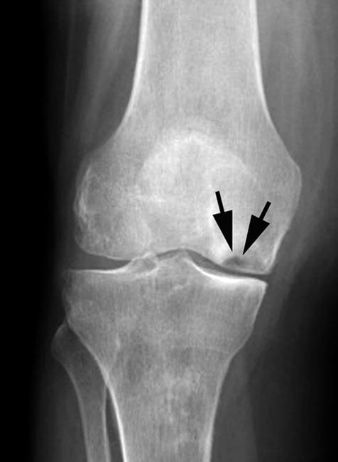
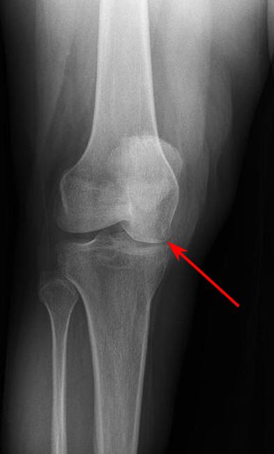
When plane radiographs were performed within a few days of the injury, it was found that he had a significant abnormality involving that knee. He suffered with so-called avascular necrosis of the medial femoral condyle. This is an area of death within the bone which forms part of the lower end of the thigh bone and would definitely have been present prior to the injury. It is possible however, although relatively unlikely, that he may have been completely unaware of the condition at a clinical level before the injury. A very careful and thorough scrutiny of his clinical records failed to reveal any evidence that he had ever complained of any problems referable to that knee joint. The benefit of the doubt therefore lay with him.
The defendants argued that the subject injury had simply unmasked this condition and had not really caused any particular damage or harm.
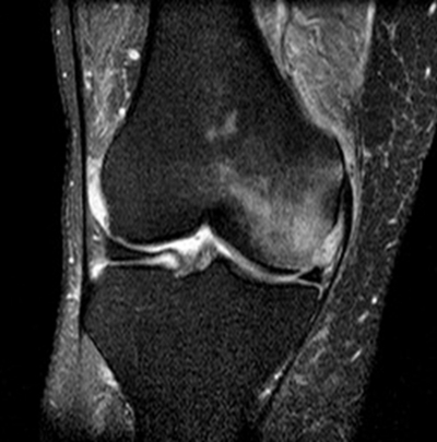
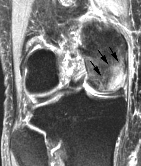
Fortuitously, he had been subjected to an MRI scan examination within three weeks of the subject accident. Not only did the MRI scan examination show these old changes of avascular necrosis involving the medial femoral condyle, but the scan also showed oedema in the bone and the soft tissues in that region. So extensive was the oedema in the soft tissues that it was reasonable to assume that quite considerable forces had been applied to the joint at the time of the subject injury and that at least some of his presentation was due to the injury specifically.
This became of considerable importance. Although he had been asymptomatic prior to the injury and it would be reasonable to assume that he may have continued in that asymptomatic state for some considerable time into the future, his clinical course was altered by the injury. Within six months, he had undergone a partial knee replacement. Unfortunately, that intervention failed and within thirteen months, he had undergone a total knee replacement. That intervention was also not particularly successful and given the fact that he was still in his fifties, it was more likely than not that he would eventually require a revision of that total knee replacement.
He had therefore gone from a state of being completely asymptomatic to a circumstance whereby he had been subjected to two major operations yet remained seriously encumbered.
He was successful in his civil suit and the quantum he received was thought by the defendant to be extremely generous. Whilst that may have been the case, had it not been for the MRI scan examination findings, I suspect that he would have received little, if anything.
In this situation, the MRI scan examination appeared to be pivotal.