Category Archives: Uncategorized

April 2018 – Case Vignette

Case Vignette

Multiple Injuries In The Lumbar Spine

The AMA 5 Guides recommend that expert reporters quantifying impairments use the so-called “Diagnosis Related Estimate Category” method for assessment.  There are several reasons for that recommendation.  One relates to the potential for unreliability when assessing ranges of motion of the lumbar spine.  The assessor may be mistaken or alternatively, the patient may be inconsistent and/or even overreactive.  The use of the Diagnosis Related Estimate Category system assists in levelling the playground for assessment.


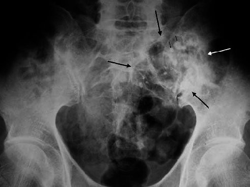
The Guides however are equally specific in their advice concerning multiple injuries in the same vertebral column segment.  That is to say, if there are two or more discrete injuries in the lumbar spine, it is not the Diagnosis Related Estimate Category that should be used but rather, the Range of Motion method.  For example, if there is a fracture of the transverse process, a fracture of a vertebral body at L3 and a fracture of a lamina at L5, it is the Range of Motion method that should be used.

Multiple lumbar vertebrae fracture

There are two steps to the Range of Motion analysis.  The first relates to measuring the losses as a result of the specific fractures according to Table 15-7. There are separate sections within that Table that make reference to fractures of posterior elements, a vertebral body, disc lesions or forward slippage of one vertebral body on the other (spondylolisthesis).

l5 s1 mri

In addition to quantifying the loss related to the two or more discrete injuries in that lumbar spine, the reporter must also measure the ranges of motion in the sagittal and coronal planes.  Rotation should also be measured, especially in the thoracic spine.


The losses measurable using Table 15-7 and the losses measurable related to the restrictions in range of motion are then combined using appropriate Combined Values Tables to yield a final functional whole person impairment.


This can lead to confusion amongst reporters.  It is confusion that causes disagreement and usually, it is disagreement that compounds the processes of mediation and Court trials.

l5-s1 xray mild

It is most useful for all involved if your expert reporter is not only familiar with disorders of the vertebral column, but also the proper use of the AMA 5 Guides.


April 2018 – General Advice

GENERAL ADVICE

Where Does Medicolegal Experience Come From?


The answer is pretty obvious.  From all sorts of places, really.


From the medical perspective, competence is directly proportional to experience.  That is not to say that the two equal each other but as medicolegal reporters age and gain experience, so do they refine their process and diminish or even eliminate obvious errors, commissions or omissions.  There is an adage that “Good medicolegal reporting comes from experience, experience comes from bad medicolegal reporting”.  It would be a very unusual medical practitioner who engages in medicolegal reporting who has not make a few mistakes.  It should be hoped that the reporter has learnt.

Nero

From the legal perspective, experience is also important.  Whilst the legal aspects of managing a claim are well outside the medicolegal reporter’s domain, so is a precise understanding of anatomy, physiology and pathology quite difficult for many lawyers.  Whilst I am surprised at just how much medicine some of my senior legal colleagues know and understand, there will always be some voids and deficits.  There is a number of relatively easily digested texts that deal with medicolegal issues and which explain some of the nuances of injury, impairment and disability.  Another useful port of call is to telephone your friendly medicolegal consultant.  This type of dialogue assists both sides.  There is no property in a witness.  There is no reason why these frank discussions cannot occur.  Provided the rules of evidence are obeyed, considerable mutual assistance can be gleaned.

Penguins

April 2018 – Lead Article

LEAD ARTICLE – APRIL 2018

How Useful Are Past Medical Records?


I usually answer “vital” but there are some obvious exceptions.


When assessing a functional loss or quantifying an impairment, and attributing that loss or a component of the loss to a compensable event, it is important to measure any loss that may have predated the subject accident.  This process of apportionment is often overlooked and can lead to unrealistic expectations and wasteful expensive litigation.


Conversely, past medical records can be misleading.  I recall appearing as an expert witness in the District Court in Mackay many years ago.  I had flown up the evening before and dined with the barrister engaged by the solicitor who had requested my attendance.  I remember the evening well.  We dined on mud crabs.

cooked mud crab (1)

The case was first on the list the following morning and we duly arrived just before 10:00am.  The barrister that was taking my evidence in chief was acting for the defendant.  The plaintiff had allegedly injured his back in a lifting incident whilst working at one of the local cane mills.  As we walked up the steps of the District Court in Victoria Street, the barrister informed me he had read somewhere in the past medical history that the plaintiff had sustained some form of back injury.  I made a mental note.


During the course of the examination in chief, the barrister with whom I had dined mentioned this past history of back problems and questioned whether or not that could have been responsible for his current circumstance rather than the accident in question.  I foolishly answered “yes”.


My response was foolish for several reasons.  The first was that I had not taken a history from the plaintiff about this particular event that had allegedly occurred prior to the subject accident, I had not read the notations to which the barrister referred and had no idea about the significance or otherwise of this antecedent problem.


The barrister undertaking the cross examination appeared to be about as naïve as me.  He did not bring it up either.

35630932 - dvd drive on laptop computer.

35630932 – dvd drive on laptop computer.


My come-uppance was brought about by the District Court judge himself.  At the conclusion of the cross examination, His Honour asked me for more detail about this past medical history and how it could have an effect upon the opinion that I had previously proffered.  He produced the documents in question and it was revealed that the patient had previously suffered with a mild episode of coccydynia.  This is a condition that affects the coccyx and not specifically the lower lumbar spine.  Apparently, the condition had arisen “out of the blue” and without any precipitating traumatic event.  There was no previous history of previous back problems and my unwise apportionment of blame was exposed for the worthlessness that it contained.  This was a most embarrassing moment.


It was one of many lessons that I have learned.  Whilst past medical history is of vital importance, it deserves to be carefully scrutinised.  Whilst it may be of some value in the final analysis, it may be totally unrelated.  I have not made that mistake again.

thumbnail_doctor showing x-ray plates to patient

Case Vignette – April 2019

CASE VIGNETTE – APRIL 2019

An Evening At The Broncos

Try to imagine a winter’s afternoon, a Friday, at about 5:30pm. I was sitting in my consulting rooms on Wickham Terrace with my back to the window. The window gave a splendid view of the city lights. Seated opposite the desk was a young couple, probably in their late thirties or early forties. They had two children, both boys, aged 11 and 8.

The wife was the patient. She gave a six weeks history of lower back pain which was almost constant, definitely disturbing her sleep and without any particular radiation into the lower limbs suggestive of a sciatica. She had been reviewed by her general practitioner on numerous occasions, had undergone a month of physiotherapy and had also attended a chiropractor.

Senior male doctor looking at worried couple. Man is consoling sick woman. They are sitting in hospital.

Clinical examination revealed some tenderness in the lower part of her lumbar spine and in particular, lateral to the sacroiliac joint. This joint is made up of the sacrum (which is part of the vertebral column) and the hemi-pelvic wing on that side.

I looked at the radiographs. Those of the lumbar spine were unremarkable. The CT scan examination that had been arranged was also within normal limits. There was no sign of any disc narrowing, disc bulging or neural compromise.

What I did see however caused considerable alarm. It was on the left side, medial to the SI joint, and involving the sacrum proper. The lesion was approximately 5cm across and it looked as though a small shark had taken a bite. This was her first presentation with breast cancer. This was a secondary deposit in the pelvis, destroying bone and heralding a premature end to her life.

thumbnail_Conventional-radiograph-of-the-pelvis-revealed-an-osteolytic-lesion-arrows-with-a

The cause for this problem was clearly outside my purview. I broke the news as gently as I could, I made several telephone calls while they sat in my office and I made arrangements for her to be seen by both a breast surgeon and an oncologist the following morning, the Saturday. It was obvious that they were grateful but more importantly, they were overwhelmed with grief.

The level of grief, its intensity and its sudden onset were all understandable. There was however a dimension that I had not expected.

The four of them (the husband and wife with the two boys) were en route to a home match of the Broncos. They were trickling down the hill towards Lang Park. They were going to stop for a bite to eat at a hamburger joint on the way. The family was very excited.

thumbnail_Broncos Rugby

Amongst the myriad of thoughts passing through the minds of this young couple was how they would go out into the waiting room and break the news to the boys. Would they hold the news until tomorrow so that the boys could enjoy the Broncos match, would they tell the boys but still go to the match anyhow and try to enjoy themselves, or would they tell the boys and simply abort the evening and go home?

To this day, I cannot quite remember what decision they made. I do remember though feeling the grief for them. I suppose there is half a dozen memories I have like this after 40 years in practice. You will understand that these memories just never fade.

Case Vignette – April 2020

CASE VIGNETTE – APRIL 2020

Fracture/Dislocations of Joints Can be Exceedingly Challenging

Consider the case of the society matron (only 61 years of age) who sustained a fracture/dislocation of her dominant thumb. The joint involved was between the first metacarpal and the proximal phalanx of the thumb. As you look at your thumb, it’s not the end joint but the one next to the end joint.

Her hand became caught in the stock as she fell whilst skiing and the force applied bent the thumb well away from the index finger. One of the bones in the joint was fractured and was quite markedly displaced.

Upon returning to Australia (from her favourite location in Aspen), the society matron underwent an operation. The Orthopaedic Surgeon reduced and fixed the fracture, and held the thumb in a splint for six weeks.

Although the fracture healed, upon removal of the splint, it was apparent that one of the ligaments that was stabilising the joint was no longer functioning satisfactorily and the thumb itself became very unstable. This is sometimes referred to as “a gamekeeper’s thumb”. In times gone by, gamekeepers used to snap the necks of pheasants and other game birds by holding each bird against the chest with one hand and pushing forcibly downwards on the neck and head to extinguish life from the captured game with the other. The force applied to that same joint in the thumb was very similar to the force that had been applied to the thumb of the society matron when she fell in Aspen.

She was eventually subjected to a second operation, with additional costs. Unfortunately, that endeavour was complicated by sepsis which necessitated hospitalisation and intravenous antibiotic therapy. Her prolonged immobility resulted in deep venous thrombi forming in one of her lower limbs, with embolisation of the clots to her lungs. As is sometimes the case, one of the emboli was of sufficient magnitude to block the bifurcation of the main pulmonary artery, resulting in sudden death.

You can imagine the anguish experienced by the family. What seemed to be a simple thumb injury whilst skiing during the Christmas holidays resulted in the loss of their wife, mother and grandmother.

The subsequent claim was successful. The Court found that had the injury been dealt with properly in the first instance, and that the ligament had been repaired as well as the fracture being internally fixed, the eventual sequence of events was much more likely than not to have been less dramatic.

Whatever your view on the matter, it is incumbent upon the Orthopaedic Surgeon to manage fracture/dislocations totally and completely, and not just the fracture in isolation.

Case Vignette – April 2021

Case Vignette – April 2021

The Wrong Pathology

I usually draw upon cases that I have seen to structure this section of The Medicolegal Mind newsletter.

On this occasion, I am sourcing a dream I had recently. I was with a group of colleagues discussing the performance of another colleague. It related to a surgical procedure performed on a hip.

It transpired that the Surgeon under investigation had performed the subject operation perfectly. The problem was that it was the wrong operation!

He had performed an iliopsoas tendon release, believing that the patient suffered with intractable pain as a result of iliopsoas tendinopathy.


The real pathology however was not related to the psoas tendon but rather, to a labral tear, detachment and chondral anomaly within the hip joint itself.

There is no real need for the reader to bother about the precise pathology. Instead, attention should be focussed upon doing the right thing at the right time, for the right reason on the right patient. Without this combination of correctness, adverse outcomes are likely to be witnessed.

Case Vignette – April 2022

Case Vignette – April 2022

How Much Is An Injury Really Worth?

I recall a youngish woman (33 years of age) who was bumped by a horse and fell gracefully onto soft lawn. The entire event was witnessed by many people.

No doubt, it was a little frightening. She also had an anxious disposition, was embarrassed and began crying inconsolably.

Everybody reasonably expected that that would be the end of the matter; but oh no, it was not! She sued the horse owner and over the ensuing three years, sought advice and treatment from almost every medical practitioner imaginable.

She was eventually awarded $630,000 by the trial Judge in her civil action undertaken here in Brisbane.

I suppose I can’t be certain that this was nothing more than a “try on” but I am genuinely suspicious.

Case Vignette – August 2018

CASE VIGNETTE – AUGUST 2018

A Simple Stumble – A Lifetime Of Misery

I recently saw a 45 year old male who sustained a knee injury whilst leaving work one afternoon.  The injury occurred in winter of 2015.  It was only 5:45pm but it was already dark.  He was leaving a donga on a work site in central Queensland.  There was only one step from the donga down to the uneven ground below but somehow, he tripped and fell.  He did not actually roll onto the ground but instead, took all of his weight on his right foot with his knee joint partially bent and his torso twisting to one side.  He experienced immediate pain.

Sep 18 CV - man about to trip on yellow metal beams

He was helped by friends, taken to a local hospital, spent the night in bed with analgesics and elevation, and was then referred to a regional centre for further assessment.  He had sustained fractures involving the upper end of his shin bone (the tibia) and both sides of the tibia that contributed to the knee joint (the tibial plateaux).  The fractures were displaced and the joint surface was no longer pristine and smooth.  The treating surgeons opened the fractures, reduced the fragments as best they could and used metal plates and screws with some biosynthetic bone graft in an effort to reconstruct the upper end of the shin bone.

As valiant as the efforts were, a so-called anatomical reconstruction could not be achieved.  Over the next 20 months he was subjected to braces, physiotherapy, crutch ambulation, analgesic ingestion and several programmes of rehabilitation.

Aug 2018 CV (a) and (b) LL films

As nature would have it, the fractures did heal but the joint surface was no longer smooth or congruous.  In addition, the soft tissues had been damaged quite severely and whilst the ligaments remained intact, the surrounding capsule became adherent to the lower thigh bone and upper shin bone.  The knee joint became stiff, painful and swollen.


Now it’s early days but clinical and radiographic examinations confirm that this knee joint is not retrievable.  He is likely to require a total knee replacement within the next couple of years.  That puts him in his late forties, and this is very early for this type of operative intervention.


When total knee replacements are performed in patients in their sixties, the arthroplasty or artificial joint has a survivorship of approximately 90% at 20 years.  That is to say, only 0.5% of these joints fail annually.  Failure is defined as the joint requiring revision or a second replacement.


The results of knee replacements in younger men are not quite as good.  His failure rate could actually be twice that.  By the time he is 70 therefore, he has a 40% chance of that joint requiring revision.  The primary replacement would have cost in the order of $50,000.  The revision will involve costs in the order of $60,000.


Apart from the knee, this poor fellow was otherwise in excellent health.  There is no reason to believe that he may not live until his mid-eighties or even longer.  In fact, his father lived to 101 and his mother, whilst dying in her early seventies, was fatally injured in a road traffic accident.  In other respects, it is probable that she would also have lived until her late nineties or even beyond.  This chap’s genetics therefore are such that he could also live at least that long.


That therefore raises the spectre of him requiring a re-revision.  Costs in the order of $70,000 may be involved.  He may have a 15%-20% chance of requiring this re-revision.


Aug 2018 CV - 4 knee films

Added to the complexities of these surgeries is the risk of complications.  Arteries or nerves may be damaged, he may suffer with deep venous thromboses or emboli travelling to the lungs, he may suffer a stroke or even a debilitating cardiac event.  The infection rate for the primary arthroplasty is only about 0.6% but with the revision and the re-revision, the infection rate may double, treble or even quadruple.


This simple fall therefore has the ability to open up a very unpleasant further 40, 50 or even 60 years.  Unless your reporting expert is aware of these scenarios, your client is at risk of being severely under-done at the time of settlement.  Choose your expert wisely.


Case Vignette – August 2019

CASE VIGNETTE – AUGUST 2019

Lovers’ Heels
How many of you have heard of this injury – Lover’s Heels?

In simplest terms, it refers to fractures of one or both of the calcanei or heel bones. It is classically sustained by a male who has been unexpectedly found in a bedroom with another man’s wife. As the husband of the household ascends the steps to the bedroom, the lover jumps from the bed, runs to the verandah and leaps over the railing. Landing on the concrete driveway three metres below gives rise to this awful injury.

man jumping off balcony

Heel fractures are particularly troublesome in orthopaedic surgery. They give rise to extreme pain and extraordinary swelling. Bed rest, elevation, ice and analgesia are the hallmarks of early treatment, especially if the fracture is extensive and comminuted (many fragments).

The calcaneus or heel bone forms a very important joint with the ankle bone or talus. This so-called “subtalar joint” allows the heel to move inwards and outwards when walking over uneven terrain. If this movement is restricted as a result of post-traumatic arthritis, walking becomes particularly arduous and almost impossible on soft sand, construction sites or through the bush.

calcaneal-fractures

Whilst these fractures can be opened surgically, pieced back together a little like Humpty Dumpty and held in position with plates, screws, wires and other devices, the joint between the talus and the calcaneus is often left irreparable.

heel fracture

The salvage operation is in the form of a subtalar fusion or arthrodesis. Arthrodeses are very important in that they convert a stiffish, painful joint into a stiff, painless joint. The afflicted individual loses the 10° or so of inversion and eversion of the heel, but also loses the pain. It is a most attractive trade-off.

My advice to men in this situation is initially to avoid it altogether but if the attraction is so great, stay in bed and just face the music. It’s likely to be less painful than the calcaneal fracture!

violins

Case Vignette – August 2020

CASE VIGNETTE – AUGUST 2020

Bad Luck or Negligence?

Pedro was a 73 year old immigrant who had previously been quite active. He played tennis, walked regularly and also rode his bike.

He recalled injuring his right hip joint whilst playing tennis a few weeks prior to his first presentation to his local medical officer. The doctor was perplexed but made arrangements for an MRI scan examination and sent Pedro off to see an Orthopaedic Surgeon.

The surgeon did see some degenerative changes on the MRI scan, but considered that a non-operative approach would be appropriate.

Pedro re-presented six months later and according to his account, the pain was really much the same. The surgeon however recommended a total hip replacement. Pedro claims that he trusted the doctor implicitly and it was for that reason that he proceeded.

Unfortunately, some technical errors occurred during the course of the hip replacement and in addition, Pedro suffered with a late septic complication.

I was asked for an opinion concerning two factors.

The first factor was whether or not the hip replacement was really indicated. This is a difficult circumstance to assess in hindsight. It is really best judged by those present at the time. It is noteworthy that the Orthopaedic Surgeon had seen him on two occasions, six months apart, and on the first occasion, recommended a non-operative approach. The plane radiographs and the MRI scan examination did demonstrate quite severe disease and Pedro was obviously in his seventies. On balance, it is more probable than not that the recommendation to proceed towards a hip replacement was reasonable.

The second issue related to the technical errors that occurred during the course of the hip replacement and whether they were related to the sepsis.

There was no link between the technical errors and the sepsis. Unfortunately, septic complications occur in just under 1% of hip replacements and despite the best of endeavours, there is little more that we can do. On perusing the notations, it was obvious that the Orthopaedic Surgeon had taken every recognised precaution in an effort to prevent the septic outcome.

The technical errors were relatively minor. The test to be applied is whether or not the performance on the part of the surgeon was equivalent to, or better than, what would be expected of an average Orthopaedic Surgeon in Australia today.

Again, on the balance of probabilities, his performance was satisfactory.

Ultimately, this has been a very unfortunate outcome for Pedro. He is now far worse off than he had been and in retrospect, would have been better avoiding surgical intervention.

I suppose you have all heard that saying “Hindsight is better than foresight by a damned sight”.