Category Archives: Uncategorized

Case Vignette – August 2021

Case Vignette – August 2021

Horses are Heavy

A 28-year-old secretary was visiting a local winery on a Sunday afternoon. By all accounts, she enjoyed herself enormously. The food was excellent, the wine flowed copiously and although she was able to walk to her car, considerable aid was required.

She demanded a detour en route to the car park. She had spied three chestnut geldings in a nearby paddock. Imbued with both liquor and love for animals, her approach was too close. The nearside front hoof of one of the big fellas trod on her sandalled toes.

Her state of inebriation was such that she probably didn’t feel a lot, although three of her digits were amputated immediately and the remaining two looked quite dusky. Over the next several weeks, one of those two toes required amputation but the fifth digit (her hallux or great toe) was saved.

Many months of rehabilitation followed and her claim included Post Traumatic Stress Disorder.

Although there was no padlock on the gate to the horses’ paddock, it was firmly closed and required two hands to disengage the catch. There was also a small sign warning guests about entering the paddock and identifying it both as private property and a container of danger.

Whilst everybody was very sympathetic, her claim was ultimately unsuccessful. It just goes to show – not all long lunches end well.

Case Vignette – December 2018

CASE VIGNETTE – DECEMBER 2018

ASSESSING SPINAL INJURIES USING THE AMA 5 GUIDES

Chapter 15 of the AMA 5 Guides explains specifically that whenever possible, the Diagnosis Related Estimate method should be employed. For example, if there is a fracture of L3 that has given rise to a loss of 30% of normal anterior body height, the assessor would refer to Table 15-3 on page 384. Fractures that give rise to a loss of 25%-50% of normal anterior body height quality for DRE Lumbar Category 3 with a range of 10%-13% of whole person function. That range allows the assessor to make some allowance for associated pain and discomfort. A patient who has sustained such a fracture but is otherwise relatively unscathed, would be assessed at the lower limit of the range and given a 10% whole person impairment rating. Conversely, another patient with that same fracture but who has near constant pain and considerable difficulties with a broad spectrum of activities of daily living may rate at the upper limit of that range and qualify for a 13% impairment assessment.

L3 fracture xray

Consider however the patient who has more than one fracture within the lumbar spine. Let’s say that there is a loss of 10% of anterior body height at L1 as a result of a compression fracture with another loss of 30% of anterior body height at L3. In this circumstance, the AMA 5 Guides instruct the assessor to use the “Range of Motion” method. This is best outlined in Table 15-7. It appears on page 404. The fracture at L1 (a loss of 10% of anterior body height) would qualify for a loss of 5% of whole person function. The fracture at L3 with a loss of 30% of anterior body height would qualify for a loss of 7% of whole person function. Those two losses would be combined to yield a loss of 12% of whole person function. In addition, the assessor must also make allowances for any restrictions in range of motion. Tables 15-8 ad 15-9 make allowances for losses of flexion, extension and lateral flexion to the right or left. This is a more complex analysis but is demanded by the AMA 5 Guides because there is more than one segment within that same region of the spine which has been injured.

lumbar fractures xray

To make it even clearer, if there was a fracture in the cervical spine, a fracture in the thoracic and a fracture in the lumbar spine, the assessor would still use the Diagnosis Related Estimate Category method and refer to Tables 15-5, 15-4 and 15-3 respectively. It is only when there is more than one fracture in the same region (cervical, thoracic or lumbar) that Table 15-7 is used.

Case Vignette – December 2019

CASE VIGNETTE – DECEMBER 2019

Even Santa Claus is Vulnerable

Folklore has it that Santa became stuck in the chimney of an old house in East Brisbane in December 2017.

There was no particular obstruction in the chimney flue itself but he was obese, his sack was heavily laden and the bells on his waist became caught on the roughened interior.

Fortunately, Santa could breathe but his cries for help were muffled and the terrified children who found him soon after dawn were understandably shocked.

The Emergency Services were able to retrieve Santa and took him to the Princess Alexandra Hospital. Whilst radiographic examinations excluded any fractures, his left lower limb had been caught in a fully flexed position and the blood supply to the muscles below his knee had been seriously curtailed for more than four hours.

It became apparent during Christmas Day that Santa had a compartment syndrome. Despite immediate decompressive fasciotomies, multiple operative procedures thereafter with skin grafting and expert care, his limb was deemed to be non-salvageable. Santa was subjected to a below knee amputation in the third week of January.

Even though this case is rare enough to be worthy of a report, the scenario is quite well recognised. Inebriated revellers can fall asleep on a toilet, fall in remote locations without assistance, or be otherwise denied prompt therapeutic intervention, resulting in significant functional losses.

Whilst Santa was eventually successful with his personal injury claim, I suspect that these events will eventually disappear from our radar. I further assume that Amazon will take over Santa’s delivery role in the future and with proper workplace health and safety initiatives, we may no longer see this type of injury.

Case Vignette – December 2020

Case Vignette – December 2020

When I was a Young Fella

When I was a year 12 high school student, I was involved in a motor cycle accident. It happened during the course of the year 12 examinations and I had to rely upon a year 11 scribe to complete the chemistry and mathematics 2 examinations on my behalf. Theoretically, I was advising him from my hospital bed but in reality, he was the star performer. I remain ever grateful to him.

I sustained fractures involving my tibia and fibula, my distal femur and my pelvis. My convalescence was complicated by a pulmonary thromboembolic event.

As is usual, a third-party personal injury claim was made and two senior Brisbane orthopaedic surgeons proffered opinions for the Court. Both surgeons accurately identified the problems that had been sustained at the time of the accident. Unfortunately, neither surgeon gave much thought to the long-term effects that I would eventually witness five or more decades later.

I have since undergone two joint replacements, a joint fusion and a spinal fusion. I have had further pulmonary thromboembolic complications.

Whilst neither orthopaedic surgeon had a crystal ball, these types of sequelae are foreseeable.

I suggest that when you are engaging an orthopaedic surgeon to report on your client, you ensure that he or she takes into account the long-term adverse effects that may accrue.

Case Vignette – December 2021

Case Vignette – December 2021

It’s All a Matter of Scale

I recently saw a young man who sustained an injury in the region of his cervical spine whilst playing touch football at an Army base in 2015. It appears as though it was a genuine injury giving rise to a discal protrusion and he was subjected to an operative discectomy.

As is sometimes the case, some of the disc material was not removed per-operatively and a further disc protrusion resulted eight or nine months later. It was confirmed with a second MRI scan. As a result, a second operation was performed.

Now, this chap was unlucky. A further recurrence occurred after a trivial incident at a restaurant and yet a third operation was required.

He was then involved in a relatively minor rear end collision. I say relatively minor because he did not lose consciousness, the rear of his seat was not broken, the damage to the vehicle was less than $1,000 and he was able to drive it from the scene of the accident for a further seven hours to a north Queensland location.

His cervical spinal symptoms however were apparently increased and six or seven months later, he was subjected to a fourth operation in the form of a cervical fusion.

His legal advisor suggested that he pursue not only the Australian Defence Force Army, but also the insurer covering the driver of the offending vehicle. It is always difficult to apportion blame accurately. In general terms however, it appeared to me that he was always destined to require that fourth operation and the rear end collision was of little or no significance.

What do you think?

Case Vignette – February 2019

CASE VIGNETTES – FEBRUARY 2019

Who Would You Get To Do Your Hip Replacement?

Lawyers typically know which fellow practitioners are good and those who are not. Whether the need is for a commercial litigator, a mediator, a criminal lawyer or a family law advocate, all of you will know just who to go to and when.

Orthopaedic Surgeons should be the same. Unfortunately, some surgeons will themselves require surgical intervention from time to time. The key question then is, who do you trust?

doc queries

question marks

This can be more difficult than you might imagine. Personality obviously has a role to play but ultimately, most of us want an expert technician with a low complication rate and with a reputation for producing results that yield a long-term survivorship or outcome.

Not many Orthopaedic Surgeons visualise other surgeons operating. Trainees are usually well-positioned because they have an opportunity to assist many Consultants in a variety of settings. Consultants themselves however are often busy at the coalface and do not spend much time in the presence of their colleagues in the operating theatre environment.

The Australian Orthopaedic Association National Joint Replacement Registry collates outcome data following hip replacements, knee replacements, shoulder replacements and knee joint anterior cruciate ligament reconstructions on an annual basis. Contribution rates by surgeons in Australia are close to 100%. Longitudinal analyses are possible and observers are therefore able to determine which surgeons are performing better than the average.

graph 2


graph

Next time you contemplate undergoing orthopaedic surgery in the form of a joint replacement, ask your surgeon how he or she fares at a national level.

Case Vignette – February 2020

CASE VIGNETTE – FEBRUARY 2020

Indolent Infection  Following Serious Orthopaedic Injuries

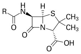
Bacterial infections continue to give rise to significant difficulties with orthopaedic surgery, whether it be in the emergency setting or at the time of elective operations. The pivotal work of Fleming and Florey bringing penicillin to market in the first two or three decades of the last century made a significant change to the outcome of surgical endeavours. Research and development by the major drug marketing companies has brought a plethora of new and different types of antibiotics into the market.

Unfortunately, our bacterial adversaries are extremely clever in the way they mutate and learn to overcome the obstacles presented by the antibiotics, and thereby thrive in the post-surgical setting. Some of the difficulty is of our own doing. There are practitioners who will use antibiotics unwisely, patients who do not always take a full course, and bacterial whose virulence sets them apart in their ability to develop resistance.

So serious is this problem that as we enter the next decade, it is likely that we will witness infections even in our own community with bacteria expressing resistance to every chemotherapeutic agent at our disposal.

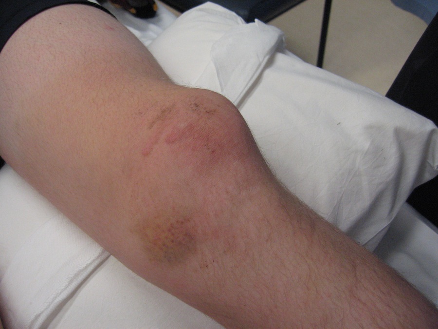
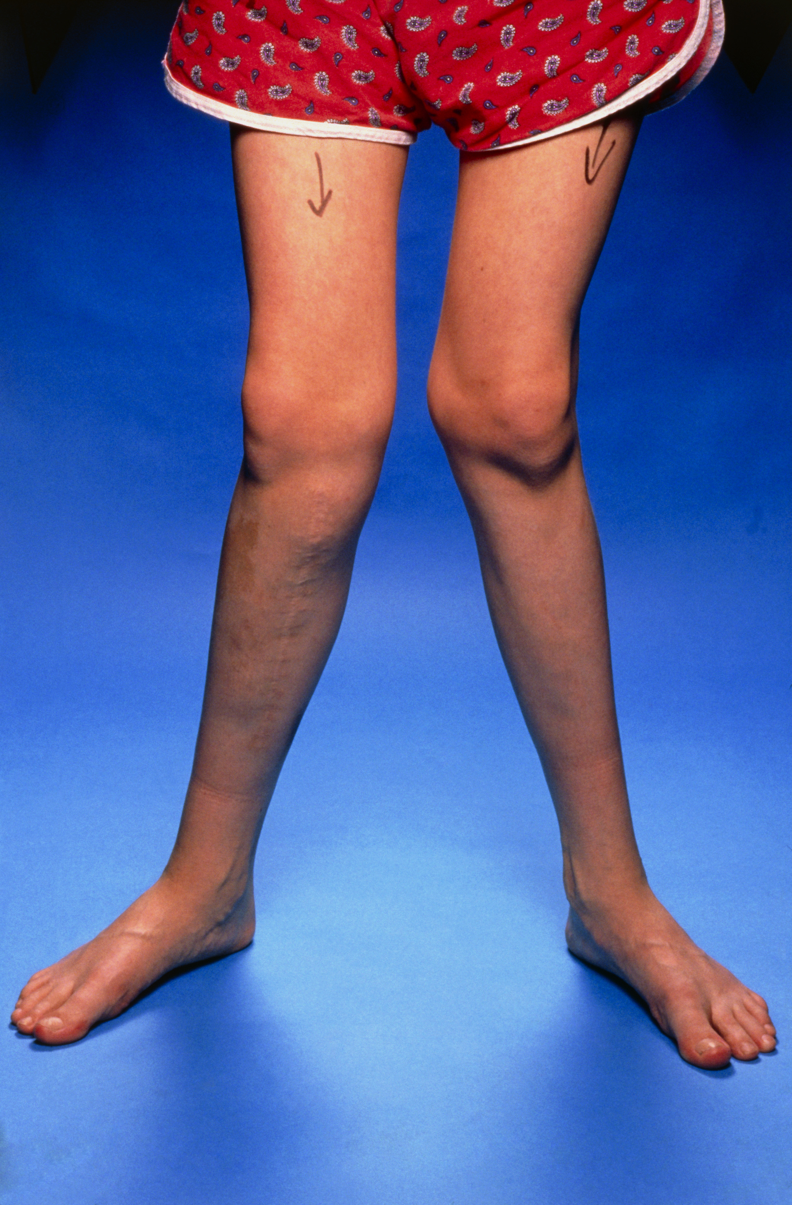
Most infections are obvious. They are accompanied by pain, redness, swelling, an increase in temperature and often a discharge (whether it be serous or pus). Other infections however are vastly more subtle and can remain indolent for weeks, months and even years.

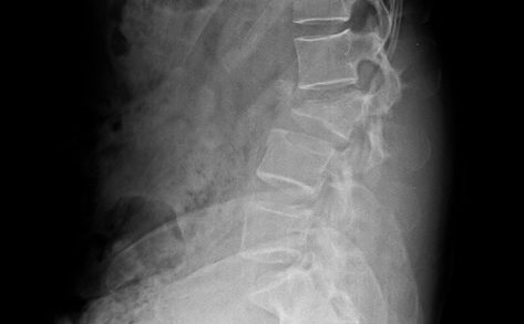
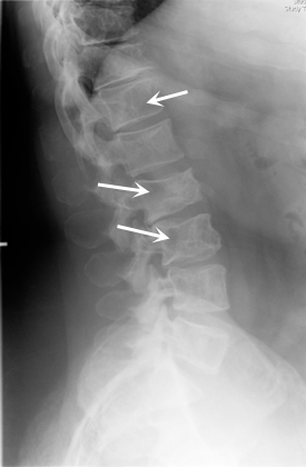
For example, it is possible to have an osteomyelitic bacterium that has lay quiescent in sequestrum in a bone for some decades. It is only when that sequestrum or local deposit is disturbed (say at the time of an injury with a fracture or at the time of undergoing a hip or knee replacement) that the bacterium is again released, prodded into action and a spectacular infectious result is witnessed.

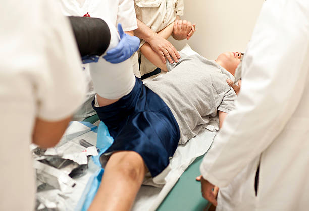
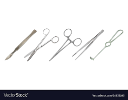
Whilst non-operative modalities such as the use of antibiotics are of great importance in fighting infections, it is the scalpel which is the most effective of all tools. Abscesses should be drained, dead and devitalised tissue debrided or removed and vascularisation of the local region maximised.

This has very special medicolegal implications. There is a risk that a patient may be deemed to have achieved the state of Maximal Medical Improvement. The condition could be thought to be both stable and stationary. A claim could therefore be closed prematurely. The recurrence of an indolent infection after claim closure could result in significant morbidity, great financial imposts and even mortality. It is worthwhile ensuring that your medicolegal expert always considers this possibility when formulating a report.

Case Vignette – February 2021

Case Vignette – February 2021

My Mate with the Medial Meniscal Tear

A mate of mine was playing touch football recently and sustained a bucket handle tear involving his medial meniscus. He is in his late forties but is otherwise relatively fit and well.

An MRI scan examination has confirmed that the bucket handle tear exists.

Whilst there is little argument that he will benefit from an arthroscopic procedure, there are two operations that could be performed.

The first is to undergo a simple resection of the bucket handle component. It can be performed as a day case, he may benefit from one or two visits to a physiotherapist, the wounds will have healed within seven to ten days, and he can effectively get on with his life. He has been rendered more vulnerable to the onset and progression of osteoarthritis in that particular compartment of the joint because of the partial meniscal loss. It may be 20, 30 or even 40 years however before the trouble becomes apparent.

The alternative approach is to perform a so-called meniscal repair. This is heavily dependent upon the tear being in the peripheral vascularised zone. If it is not there, and if there is not sufficient blood supply, healing will not occur. Assuming however that it is in the peripheral zone and that it is well vascularised, a repair might be successful. Not all repairs however are successful. It is possible for example that my mate may have to undergo another operation. At the second sitting, maybe that meniscal segment will be removed after all. What could have been a week or two of discomfort can then be extended out to being many months of difficulty, with grossly escalating costs.

Obviously, there is no single answer. Every patient should be treated on his or her merits. It occurs to me however that there are times in medicine when we are driven by altruism to go just a little bit too far. What do you think?

Case Vignette – February 2022

Case Vignette – February 2022

The Features Some Plaintiffs Suffer

I saw a fellow recently who had been working in western Queensland as a helicopter musterer for about 11 years. He had obviously started early because he was only 30 years of age when the accident occurred. He had been executing hair-raising manoeuvres through a heavily wooded forest in search of the final beast or two. He thought he would sleep better at night if the herd was complete.

The inevitable happened and the helicopter did crash. Fortunately, there was no fire although had there been, the smoke may have brought his helpers to his side more rapidly.

Instead, he sustained a severe thoracic spinal injury and was rendered immediately and completely paraplegic. He was able to use his upper limbs to drag himself from the wreckage and shelter beneath a large ironbark tree. The accident occurred on dusk and the night brought heavy rains and freezing cold, since it was July.

He realised the severity of his injury and genuinely thought that he was going to die in that remote location. He nearly did. Two full days elapsed before the rescue team found him.

By then, his lower half was covered in excrement, he had almost been eaten alive by ants and he was delirious with hunger and thirst.

I met him for his medicolegal examination four years later. His face was suntanned, his upper limbs were strong and equally brown and his wife of seven years accompanied him. Although he was confined to his wheelchair, it did seem that most other facets of his life were back on track.

So harrowing was his experience, I encouraged him to write a book. I genuinely hope he does.

Case Vignette – January 2019

CASE VIGNETTE – JANUARY 2019
PRE-EXISTENT ABNORMALITIES CAN BE MADE WORSE
A 25 year old male cyclist was dislodged from his bike whilst riding out towards the Brisbane Airport. He landed awkwardly and sustained a dislocation of his right patella. This is where the knee cap moves to the outside of the lower end of the thigh bone. It can be particularly painful, sometimes spontaneously reduces at the scene but sometimes also required transfer to a hospital with a closed reduction being effected under a Neurolept anaesthetic. He did require that form of closed reduction.

patella dislocation image

Interestingly, the knee joint was not normal before this bicycle accident. The young man suffered with what is called generalised ligamentous laxity. It is sometimes called “double-jointedness” but really, it refers to the fact that ligaments which stabilise joints are relatively long or lax, that the range of motion in almost every joint in the body is more than you might normally detect and does predispose the individual to joint instabilities or so-called subluxations or even dislocations. In addition, his knee cap was riding higher than normal and well outside the groove on the lower end of the thigh bone that usually assists with stabilisation. The knee cap was also smaller than normal and the stabilising groove on the femur was shallower than normal. In essence therefore, he had a number of features that predisposed him to dislocation.

genu valgum picture

Despite these predisposing factors, and they were quite powerful, he had never suffered with any such dislocation. It was not until the bicycle accident occurred that the patellofemoral joint became so disordered. Had it not been for the accident, he may never have suffered with a dislocation.

Quantifying his impairment and his disability was theoretically complicated by these predisposing factors, but should not have been. Regardless or otherwise of the predisposing factors, he was subjected to a single specific traumatic event and did suffer with an identifiable injury which was compensable. This is exactly what the Court ultimately found.